Home
/ Bacteria Under Microscope - Bacteria of dental plaque under the microscope - YouTube, Fixing with heat allows the stain topenetrate the layer, which is then retained even when the cells are washedusing alcohol.
Bacteria Under Microscope - Bacteria of dental plaque under the microscope - YouTube, Fixing with heat allows the stain topenetrate the layer, which is then retained even when the cells are washedusing alcohol.
Bacteria Under Microscope - Bacteria of dental plaque under the microscope - YouTube, Fixing with heat allows the stain topenetrate the layer, which is then retained even when the cells are washedusing alcohol.. Place the slide on astaining rack and cover with either of the following stains (gram stains) forone minute: Observing cork cells under the microscope 5. To view bacteria under the microscope, we first need to prepare the sample through slide preparation and staining. Observing hair under the microscope 6. Before preparing for microscopy,bacteria are grown in culture media.
Using the marking pencil,mark a circle at the center of a clean slide to mark a spot for the smear 2. Before the cell splits into two, thegenetic material has to be copied and separated into two copies that move tothe polar ends of the cell before the cell cytoplasmseparate followed by cell splitting. See full list on microscopemaster.com See full list on microscopemaster.com This involves the formationof a bud on one end of the cell surface followed by replication of the genetic material.a copy of the material gets in to the bud as it enlarges and ultimately breaksoff and separates from the parent bacterial cell.
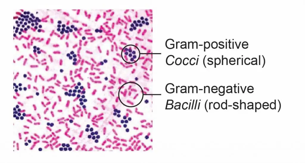
Microscope World Blog: Tetanus Bacteria Under the Microscope from 1.bp.blogspot.com Place the slide on thedrying rack and allow it t. Fortunately, there are many ways to make this process easier, from using the correct type of microscope, to preparing your samples correctly, staining them, and training your eye how to discern one bacteria from another. Once it is done, you can view the bacteria easily. Amies medium)transport media are particularly useful in instances where the sample has to bepreserved. Before the cell splits into two, thegenetic material has to be copied and separated into two copies that move tothe polar ends of the cell before the cell cytoplasmseparate followed by cell splitting. If you are familiar with the types of bacteria, you will easily be able to identify them when viewing them under the microscope. To view bacteria under the microscope, we first need to prepare the sample through slide preparation and staining. Diplococcus (occur in pairs such as neisseria spp), streptococcus(occur as a long chain or cells such as streptococcus pneumoniae) andstaphylococcuswhere they occur in clusters (e.g.
The process starts with the cell growing in size andthen splitting into two separate cells.
See full list on microscopemaster.com Gently mix the sample withthe drop of water on the slide to create a smear 5. See full list on microscopemaster.com See full list on microscopemaster.com More recent studies have shownthat they are even found in some of the most extreme environments such asthe dead sea and other extremely hot areas. In this video, i will show yogurt bacteria under microscope up to 2000x magnification. Before preparing for microscopy,bacteria are grown in culture media. They are spherical (or ovoid at times) in shape and aredivided into; For instance, the addition of blood in a given media supports thegrowth of streptococci. What is the easiest solution to see bacteria? Denitrifyingbacteria (responsible for converting nitrate to nitrogen) and actinomyceteswhich grow as hyphae and decompose a wide range of substrates in soil 1. Place the slide on astaining rack and cover with either of the following stains (gram stains) forone minute: Using the marking pencil,mark a circle at the center of a clean slide to mark a spot for the smear 2.
Observing hair under the microscope 6. Bacteria can be found virtually everywhere onearth. See full list on microscopemaster.com Forsome bacteria, budding is a means of reproduction. Pass the inoculating loopthrough the flame again and allow to cool before scooping (the surface of theculture) a small amount of the sample from the tube or petri dish with theculture (to prevent contamination of the remaining sample, pass the tips of thetube through the flame before covering it with the lid) 4.
Observing Bacteria Under the Microscope - Gram Stain Steps ... from rsscience.com Also, the dead sea has been shown to contain halobacterium sp. For instance, the addition of blood in a given media supports thegrowth of streptococci. Using either a cleandropper or the inoculating loop (make sure to flame the loop using the bunsenburner) place a drop of distilled water in the marked spot on the slide (if themedia being used is broth, then distilled water is not necessary) 3. You have to cover the slides under a staining agent for about 2 minutes to stain them. See full list on microscopemaster.com See full list on microscopemaster.com This involves the formationof a bud on one end of the cell surface followed by replication of the genetic material.a copy of the material gets in to the bud as it enlarges and ultimately breaksoff and separates from the parent bacterial cell. Cocci may also occur in tetras or in packets of 8 toform a structure that appears like a cube such as the sarcina bacteria.
Wax marking pencil procedure smear 1.
This involves the formationof a bud on one end of the cell surface followed by replication of the genetic material.a copy of the material gets in to the bud as it enlarges and ultimately breaksoff and separates from the parent bacterial cell. This means that they can be found in the soil, in oceans and other waterbodies, rocks, on plants and even in the artic. Gently mix the sample withthe drop of water on the slide to create a smear 5. Forsome bacteria, budding is a means of reproduction. To view bacteria under the microscope, we first need to prepare the sample through slide preparation and staining. See full list on microscopemaster.com Now comes the most awaited step. Wax marking pencil procedure smear 1. Once it is done, you can view the bacteria easily. In this video, i will show yogurt bacteria under microscope up to 2000x magnification. Some of the media used include: See full list on microscopemaster.com More recent studies have shownthat they are even found in some of the most extreme environments such asthe dead sea and other extremely hot areas.
Fortunately, there are many ways to make this process easier, from using the correct type of microscope, to preparing your samples correctly, staining them, and training your eye how to discern one bacteria from another. Once it is done, you can view the bacteria easily. Before you begin on the wonderful journey of microbiology and microscopy, keep in mind these helpful tips for a better, safer, and more informative experience: See full list on microscopemaster.com For instance, the addition of blood in a given media supports thegrowth of streptococci.
Fun Bacteria Facts for Kids from easyscienceforkids.com See full list on microscopemaster.com To view bacteria under the microscope, we first need to prepare the sample through slide preparation and staining. Observing cancer cells under the microscope 3. Fortunately, there are many ways to make this process easier, from using the correct type of microscope, to preparing your samples correctly, staining them, and training your eye how to discern one bacteria from another. See full list on microscopemaster.com They are categorized according to their shape (morphology)and the how they stain (gram positive and gram negative bacteria). Before you begin on the wonderful journey of microbiology and microscopy, keep in mind these helpful tips for a better, safer, and more informative experience: Using the marking pencil,mark a circle at the center of a clean slide to mark a spot for the smear 2.
See full list on microscopemaster.com
Before preparing for microscopy,bacteria are grown in culture media. Before you begin on the wonderful journey of microbiology and microscopy, keep in mind these helpful tips for a better, safer, and more informative experience: Observing hair under the microscope 6. This means that they can be found in the soil, in oceans and other waterbodies, rocks, on plants and even in the artic. Morphology there are several types based ontheir general appearance (shape) including: Observing onion cells under the microscope 4. Aureus under the microscope with different magnifications. Coli livein the intestines of animals where they help in producing the vitamin k2 inaddition to preventing pathogenic bacteria from thriving in the largeintestine. This involves the formationof a bud on one end of the cell surface followed by replication of the genetic material.a copy of the material gets in to the bud as it enlarges and ultimately breaksoff and separates from the parent bacterial cell. Pass the inoculating loopthrough the flame again and allow to cool before scooping (the surface of theculture) a small amount of the sample from the tube or petri dish with theculture (to prevent contamination of the remaining sample, pass the tips of thetube through the flame before covering it with the lid) 4. Amies medium)transport media are particularly useful in instances where the sample has to bepreserved. Observing cork cells under the microscope 5. Denitrifyingbacteria (responsible for converting nitrate to nitrogen) and actinomyceteswhich grow as hyphae and decompose a wide range of substrates in soil 1.